
Rare immune cells found at strikingly high levels in people who live beyond 110 reveal an unusual pattern of clonal expansion, differentiation and functional flexibility.
Study: CD4 CTLs in supercentenarians: Signs of adaptive expansion in healthy aging. Image Credit: Sean Pavone / Shutterstock
In a recent study published in the journal Cell Reports, researchers characterized CD4 cytotoxic T lymphocytes (CD4 CTLs) in supercentenarians.
Aging is marked by a gradual functional decline, driven by cellular and molecular alterations, including cellular senescence, DNA mutations, and mitochondrial dysfunction. Supercentenarians, people aged 110 or older, provide a model of healthy aging, attaining longevity while delaying or avoiding age-related diseases, e.g., cancer and cardiovascular disease. Their epigenomic, immune, and cardiovascular profiles appear younger for their age.
The authors previously reported an increased frequency of CD4 CTLs in supercentenarians. These cells are scarce in peripheral blood under normal conditions but have been detected during viral infections, including severe acute respiratory syndrome coronavirus 2 (SARS-CoV-2) infection. Recent studies report that CD4 CTLs can kill tumor cells across cancers, including colorectal cancer and melanoma. Nonetheless, the role of CD4 CTLs in healthy aging remains unknown.
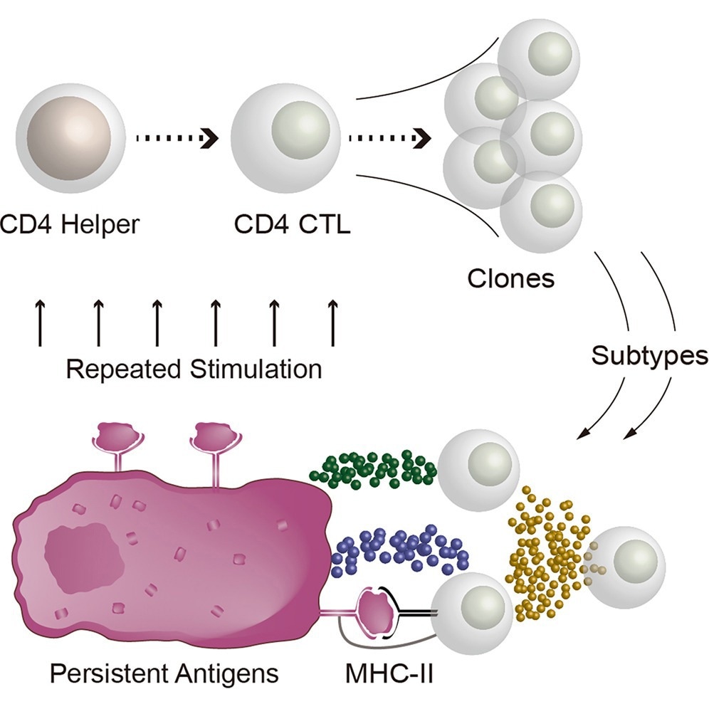
Graphical abstract illustrating the proposed expansion and diversification of CD4 cytotoxic T lymphocytes (CD4 CTLs) following repeated stimulation by persistent MHC class II-presented antigens, with clonally expanded cells adopting distinct functional subtypes.
The study and findings
In the present study, researchers characterized CD4 CTLs across older age groups, with a focus on supercentenarians. First, they generated single-cell profiles of surface proteins, transcriptomes, and T cell receptor (TCR) sequences from T cells of eight individuals aged 70–90 years, 10 centenarians, and 10 supercentenarians. In total, 43,584 T cells were collected and stratified into 13 clusters, with γδ, CD8, and CD4 T cells as the major classes.
CD4 CTLs were identified from the CD4 clusters based on the co-expression of cytotoxic genes (granzyme H [GZMH] and granzyme B [GZMB]) and CD4. CD4 CTLs became increasingly abundant across the three age groups; the median CD4 CTL proportions were 4% in non-centenarians, 9.6% in centenarians, and 17.6% in supercentenarians. Notably, one clinically healthy non-centenarian had the highest proportion of CD4 CTLs. Model-based analysis of public single-cell datasets covering more than 1,500 samples across the human lifespan further indicated that CD4 CTLs remained relatively uncommon at younger ages but increased markedly in abundance and variability at extreme old age.
The team then investigated the apparent transition of CD4 helper cells toward a cytotoxic phenotype. CD4 CTLs were marked by the loss of CD27 and CD28, distinguishing them from the remaining CD4 clusters. CD4 CTLs exhibited a distinctive phenotype of CD45RO+, CD45RA−, C-C motif chemokine receptor 7-negative (CCR7−), programmed cell death 1-negative (PD-1−), and CD95mid, indicative of advanced differentiation with no signs of exhaustion.
Transcriptional analysis further corroborated this phenotype, revealing low expression of genes associated with exhaustion, such as lymphocyte-activating 3 (LAG3) and CTL-associated protein 4 (CTLA4). A CD27−CD28+ population, comprising 10.5% of CD4 T cells, was identified as a potential transitional state between CD4 helper cells and CTLs, consistent with CD27 being lost before CD28 during this transition. The researchers then focused on TCR repertoires of CD4 CTLs, characterizing their receptor composition and clonal structure.
Clonal expansion of CD4 CTLs was detected in all samples. Further, dual TCR expression, i.e., the expression of two alpha or beta chains in a single T cell, was observed in five top clones, although dual TCRs are not unique to CD4 CTLs, and their functional significance remains uncertain. The TCRγ gene was expressed in CD4 CTLs at about half the level observed in γδ T cells. The team subsequently explored potential antigenic targets for CD4 CTLs in public TCR databases by analyzing complementarity-determining region 3β (CDR3β) sequences from top clones.
CDR3β sequences matched those of T cells expanded in samples from non-small cell lung cancer, breast cancer, and hepatocellular carcinoma. However, the matches were based on CDR3β sequences alone and do not establish that the T cells recognize the same antigens. A secondary single-cell analysis was then performed using two samples from each age group to gain insights into CD4 CTL function. Cells were split into two sets: unstimulated controls and cells stimulated ex vivo with PMA and ionomycin. Transcriptomes, TCRs, and surface proteins were profiled.
Cells were classified into eight clusters, grouped into two major classes: activated (CD69+) and quiescent (CD69−) states. These two classes contained four clusters each: CD8 T cells, CD4 naïve cells, CD4 helper cells, and CD4 CTLs. The intermediate CD27−CD28+ population was identified in both quiescent and activated states. Perforin 1 (PRF1) and GZMB were upregulated in activated cells, while GZMH and GZMA were downregulated.
Activated cells showed elevated expression of the inflammatory cytokines tumor necrosis factor (TNF)-α and interferon (IFN)-γ. A subclustering analysis of activated cells using interleukin (IL) genes revealed eight clusters, each with distinct IL expression patterns. One cluster exhibited high levels of IL-13 and IL-4, while another cluster had high levels of IL-21. Despite the differences in specific ILs, all clusters had high levels of IFN-γ and TNF-α. Highly expanded TCR clones were distributed across several cytokine-defined subgroups, suggesting that cells within the same clone can adopt different cytokine profiles after activation.
Conclusions
In summary, CD4 CTLs increase in later stages of aging, with substantial clonal expansion observed across all age groups. Some top clones exhibited dual TCR expression, while CD4 CTLs also showed the non-canonical activation of TCRγ genes. Top TCR clones, even from healthy supercentenarians, matched CDR3β sequences from cancer patients, but these overlaps do not demonstrate shared antigen specificity. Overall, CD4 CTLs may expand and diversify as an adaptive response to persistent antigens, potentially contributing to longevity and cancer suppression.
The authors caution, however, that the study did not directly establish the cells’ functions in vivo, focused on circulating rather than tissue-resident T cells, and relied on ex vivo stimulation in a relatively small cohort. CD4 CTL accumulation has also been linked to inflammatory and autoimmune pathology in other settings, suggesting that these cells may act as a double-edged sword during aging.