
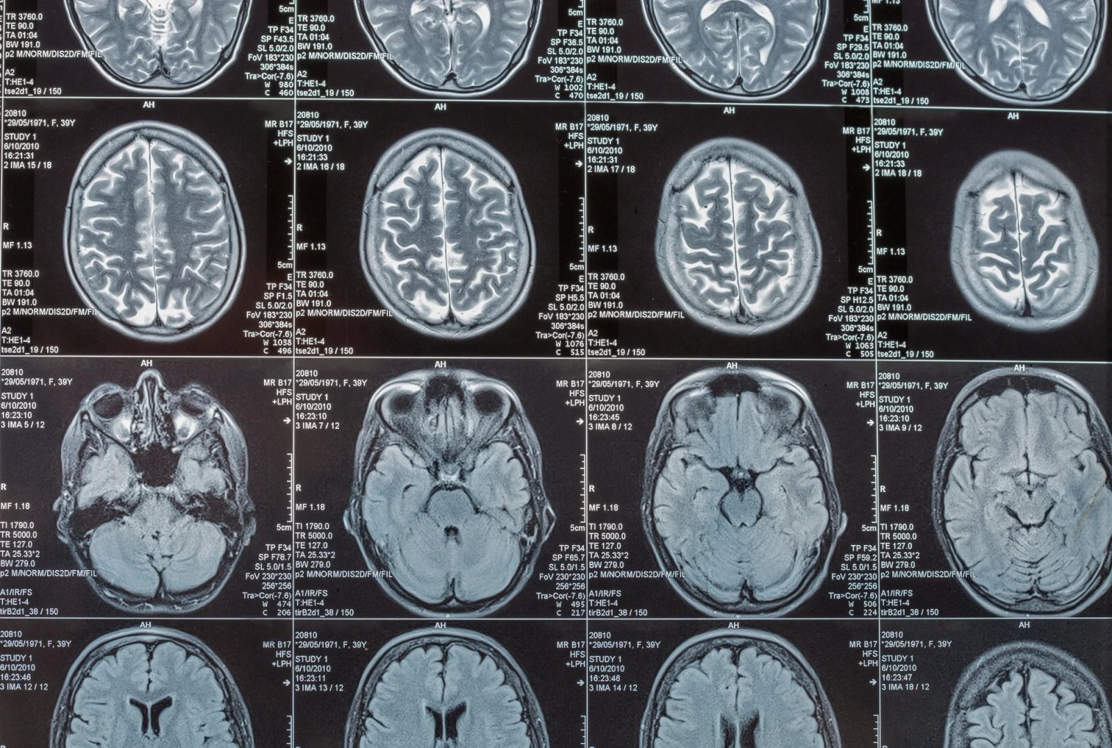
With more than 6,200 brain scans spanning healthy aging to symptomatic Alzheimer’s disease, a major research resource offers scientists a closer look at how changes in amyloid, tau and brain structure relate to cognitive health.
Study: Exploring healthy aging through preclinical Alzheimer disease in the Knight Alzheimer Research Imaging dataset. Image credit: Sergiy Palamarchuk/Shutterstock.com
A new paper published in the journal Nature Communications describes the Knight Alzheimer Research Imaging (KARI) dataset. This large, longitudinal, multimodal resource covers the spectrum of Alzheimer disease from healthy aging and preclinical AD to symptomatic disease. It identifies associations between brain imaging measures and clinical and cognitive status.
Studying the earliest brain changes linked to Alzheimer’s
Alzheimer disease (AD) is a serious worldwide public health challenge, given its high prevalence, severe and progressive clinical profile, and the lack of curative therapy. AD has a prolonged preclinical period, with amyloid and tau pathology often accumulating years or decades before noticeable cognitive symptoms. This is the period when intervention may be most useful.
Understanding this preclinical period requires multimodal longitudinal datasets: information collected from asymptomatic individuals over long follow-up periods with simultaneous assessment of molecular pathology, brain structure, cognition, and other factors. The Knight Alzheimer Research Imaging (KARI) dataset was developed by the Knight Alzheimer Disease Research Center at Washington University in St. Louis for this purpose.
The dataset’s advantages include deep phenotyping and uniform data collection standards, as it is based at a single institution. This helps maintain data quality over time, though some hardware variation is inevitable.
The present study describes the dataset and examines how its imaging biomarkers relate to age, clinical impairment, and cognitive performance. It aims to provide researchers with “the necessary context and quality-assurance metrics to utilize this expansive resource for their own investigations.”
The KARI dataset
The dataset contained 1,645 participants aged 42–97 years, from whom 6,217 neuroimaging acquisitions were obtained over the study’s data collection period. More than 800 participants had longitudinal imaging, with some followed for more than 15 years.
The KARI resource is designed to capture several aspects of brain aging simultaneously. The dataset includes structural, functional, and diffusion MRI, as well as PET imaging for amyloid and tau. These imaging data are accompanied by clinical and cognitive assessments, genetic information, fluid biomarkers, and other demographic information.
The dataset presents both raw imaging data and processed measures, including anatomical segmentations and quantified biomarkers, to qualified researchers.
A multimodal view of aging and Alzheimer disease
Most participants in the study had no signs of cognitive impairment, making the dataset particularly valuable for investigating the early, symptom-free stages of Alzheimer’s disease.
The researchers used several types of MRI scans to examine brain structure, function, and changes in brain tissue. They also used PET scans to measure the buildup of amyloid and tau, two proteins associated with Alzheimer’s disease.
The dataset included 3,385 MRI scanning sessions involving 1,571 participants, of whom 801 underwent scans at multiple time points.
For amyloid PET imaging, researchers collected 2,684 scans from 1,370 participants, including 632 who underwent repeated scans. Tau PET imaging included 1,113 scans from 826 participants, with 228 having follow-up scans.
Importantly, 792 participants underwent all three types of brain imaging: MRI, amyloid PET, and tau PET. Another 514 had both MRI and amyloid PET scans. This combination allows researchers to investigate how changes in brain structure relate to the accumulation of Alzheimer’s-associated proteins in the same individuals.
Clinical assessments were also available for 1,643 participants within two years of their brain scans. Of these, 1,132 maintained the same Clinical Dementia Rating (CDR) score across repeated assessments, while 347 had only one assessment.
The CDR scale measures the severity of cognitive and functional impairment, with a score of 0 indicating no impairment and higher scores indicating greater impairment. Most participants (1,120) had a maximum score of 0, while 362 reached 0.5, indicating very mild impairment. Another 150 reached a score of 1, indicating mild dementia, and 11 reached a score of 2, indicating moderate dementia.
Overall, the dataset includes a large proportion of people without clinical impairment, providing researchers with an opportunity to study brain changes that may occur before Alzheimer’s symptoms become apparent.
Age and clinical differences across the cohort
The mean age was 71.9 years; those with CDR > 0 were 75.7 years old on average, versus 70 years for those with CDR = 0. Thus, those enrolled with impaired cognition (CDR>0) are significantly older than the unimpaired.
The unimpaired group had more years of education and a higher proportion of women than the impaired group. Apolipoprotein E (APOE) ε4 carriers comprised 42% of the cohort, making up 56% of the impaired versus 35.5% of the unimpaired group.
Cognitive performance related to Alzheimer-related brain changes
The authors examined relationships between imaging biomarkers and the Knight Preclinical Alzheimer Cognitive Composite (Knight PACC), while adjusting for age, education, sex, and APOE ε4 carrier status.
Among 1,467 participants with MRI data, greater cortical thickness in an AD-sensitive cortical signature was associated with better cognitive performance. The partial correlation between cortical signature and PACC was 0.36.
Conversely, among 1,293 participants with amyloid PET, higher amyloid burden measured using the Centiloid scale was associated with poorer cognitive performance, with a partial correlation of −0.33.
The relationship was also evident for tau. Among 684 participants with tau PET, greater tau burden was associated with poorer PACC performance, with a partial correlation of −0.40. Thus, all three principal imaging measures showed a consistent pattern: more cortical thinning, more amyloid, and more tau were each associated with poorer cognitive performance. The associations did not significantly differ by CDR stage.
These associations remained after accounting for several important demographic and genetic factors.
Brain imaging markers vary with clinical impairment
Cortical signature decreased with increasing CDR and with increasing age. However, the decrease with age was substantially smaller in individuals with CDR 1.
Similarly, amyloid burden by the Centiloid scale increased with increasing CDR and with age, though this result may be less reliable due to the small sample size. Again, the age increase was weaker among those with CDR 1 than among those with CDR 0.
The study also examined the tau index, which reflects early tau accumulation in four brain regions. They found age-related and CDR-related increases in the index, with the effect of age being weakened in all impaired groups.
The investigators also examined tau spatial spread (TSS), a measure of how extensively tau pathology is distributed across the brain. TSS was substantially greater in impaired participants compared to unimpaired participants, and also increased with age. However, the effect of age varied with CDR.
The TSS difference increased progressively across clinical stages, from a mean of approximately 0.052 in CDR 0 to 0.682 in CDR 2.
These findings illustrate how the dataset can be used to examine not simply whether pathology is present, but how its spatial distribution differs across clinical stages, potentially informing future studies of disease progression.
White matter hyperintensities
The resource is not restricted to amyloid and tau. It also contains information relevant to vascular and mixed brain pathology, including white matter hyperintensity (WMH) volume, cerebral microbleeds and cerebral infarcts.
Radiological WMH severity (leukoaraiosis) increased with age and impairment, though the relationship with age may vary with disease severity. Cerebral microbleeds were more common among impaired participants, after accounting for age.
Cerebral infarcts, in contrast, did not show the same significant relationship with clinical status, although the sample was extremely small. These data allow researchers to investigate how vascular abnormalities coexist with or potentially contribute to neurodegenerative pathology.
Biological staging of preclinical disease
The authors also demonstrate the potential of the dataset for classifying AD according to biological changes in the brain. They applied an existing four-stage framework based on quantitative amyloid and tau biomarkers to categorize imaging sessions according to patterns of protein accumulation.
This included 656 participants with T1w MRI, amyloid-PET, and tau-PET performed within one year of each other. They underwent 812 sessions in total, of which 547 were amyloid-negative for the whole brain. Of the remaining 265 amyloid-positive sessions, 144 sessions were classified as stage A, 44 as stage B, 29 as stage C, and 43 as stage D. Five sessions were unclassified.
The investigators also examined a ‘healthy aging’ subset of 434 participants with 591 matched sessions who were negative for both amyloid- and tau-PET. They found that 372 participants across 456 sessions were unimpaired, with CDR 0 at the latest assessment.
They then looked at participants with MRI and amyloid-PET data, without requiring tau-PET. There were 647 participants with 1,248 matched imaging sessions who met the study’s criteria for remaining clinically unimpaired (CDR 0). Of these, 301 participants had data from two or more time points during the study.
This is particularly relevant to the concept of healthy aging versus preclinical AD. A person can be cognitively normal despite showing amyloid or tau abnormalities. This makes chronological age and clinical status alone inadequate to predict AD pathology in the brain. Datasets like this one allow overtly healthy individuals to be followed to identify specific brain, cognitive, or biological features that distinguish healthy aging from preclinical disease.
Strengths and limitations
The major strength of KARI is the combination of longitudinal follow-up, multimodal imaging, and deep clinical assessment. This helps identify trajectories rather than relying entirely on cross-sectional comparisons. It also helps to uncover associations between different AD components and biomarkers and the evolution of such relationships over time.
However, the study has several limitations. The imaging data were collected over many years using different scanners, field strengths, and PET tracers, leading to heterogeneity. The dataset was not completely harmonized, a deliberate choice that means researchers need to account for technical differences in their analyses.
The cohort has a relatively high educational level and is predominantly White in racial/ethnic composition, limiting generalizability. It also includes many cognitively unimpaired individuals, some of whom have Alzheimer’s-related brain pathology, meaning the findings may not fully reflect disease patterns in the wider population.
New imaging resource supports studies of healthy brain aging
Overall, the study demonstrates the value of the KARI database. Its longitudinal nature provides opportunities to investigate healthy aging, preclinical AD, and symptomatic disease through multiple disease markers and components.
It captures biological changes across the AD spectrum, as reflected in associations among measures such as cortical thinning, amyloid, tau, cognitive performance, and clinical impairment.
By combining multimodal imaging with long-term clinical follow-up, KARI could support future investigations into disease mechanisms, biomarker validation, and earlier detection, although findings will need to be confirmed in more diverse populations.
“The KARI dataset provides a powerful foundation for hypothesis generation, biomarker validation, and refinement of disease models.”
Journal reference:
-
Hoagey, D. A., McKay, N. S., Benzinger, T. L. S., et al. (2026). Exploring healthy aging through preclinical Alzheimer disease in the Knight Alzheimer Research Imaging dataset. Nature Communications. DOI: https://doi.org/10.1038/s41467-026-78439-3. https://www.nature.com/articles/s41467-026-78439-3